A Homeopathic Medicine Case Series Report
by Edward Kondrot, MD
Abstract: Conventional, allopathic treatments for age-related macular degeneration (ARMD), the most common cause of irreversible blindness in people over 65 years of age, are fraught with limitations and toxic side effects. Homeopathy, by contrast, offers a safe and effective treatment for this condition. The following three cases exemplify the unique way that homeopathy treats the individual with ARMD rather than just the condition itself; each patient required a completely different homeopathic medicine according to the Law of Similars.
Keywords: age-related macular degeneration (ARMD), dry and wet types; pharmaceutical and surgical side effects, homeopathic
treatment of; Calcarea arsenicosum, Sepia, Staphysagria.
Introduction
Age-Related Macular Degeneration (ARMD) is the leading cause of visual disability in the industrialized world and the third leading cause globally. Approximately 11 million individuals are affected with ARMD in the United States alone, with a global prevalence of 170 million. Aging is the greatest risk factor; therefore, the prevalence of ARMD in the U.S. is anticipated to increase to 22 million by the year 2050, while the global prevalence is expected to increase to 288 million by the year 2040.(1)
ARMD, the dry type, accounts for 85-90% percent of individuals with ARMD, which tends to progress more slowly than the wet type. It affects the central area of the retina called the macula that is responsible for sharp, central (high resolution) vision. The dry or areolar type consists of degeneration of the retinal pigment cells, resulting in drusen—small wart-like growths—and hyper- and hypopigmented areas in the retina, with loss of rods and cones and generalized atrophy. The condition eventually leads to fluid accumulation, hemorrhage, and scar tissue but rarely produces total blindness because the peripheral vision is preserved. The disease commonly occurs in individuals older than 65 years, but several hereditary conditions such as Juvenile Macular Degeneration (Stargardt Disease, Best Vitelliform Macular Dystrophy, Congenital Rod Cone Dystrophy) can lead to this disorder at a much earlier age.
The wet type of macular degeneration refers to the advanced neovascular (or exudative) stage of the disease, which presents a more rapid loss of vision relative to the dry type. Neovascular or “wet” ARMD arises from the growth of abnormal blood vessels from the choroid into the normally avascular sub-retinal pigment epithelium (RPE) and sub-retinal regions. Although neovascular or “wet” ARMD represents a small proportion (10-15%) of total ARMD cases, it accounts for approximately 90% of all cases of severe vision loss from the disease.(1)
There are currently no effective conventional treatments for the dry form of macular degeneration and pharmaceutical treatments for the wet form are limited by their toxicity and side effects. (see Discussion). This makes the acceptance of natural, overall health-improving therapies, such as homeopathic medicine, even more compelling. The following are three such cases.
Case 1
DS, a 76-year-old female
This patient had a prominent look of suffering on her face during the interview. She had had a recent hypertensive crisis—blood pressure 220/146, and was currently on anti-hypertensive medications.
DS was very anxious that her vision be restored. She had had over ten laser treatments in both eyes. She was worried about the future and what would happen. She wanted more information so she could do “whatever will help me.” She said, “I am a skeptical person.”
DS slept with the light on all night. She had been robbed as a child and she still had a fear of robbers. She was afraid that a man would come up through a trap door. Her parents’ home was a large fourteen room house. She and her sister would take turns staying up at night. She had a fear that someone was in the room. DS was on guard all the time. She slept on her left side facing the door. She also had a small “ladies” gun which was loaded. She slept for a couple of hours and then woke at 2:00 to 3:00 AM. The fears were worse when she was alone. She stayed up all night when her husband was in the hospital. She said that she could solve this problem if she had a dog. The dog could be on guard instead of her.
Physical exam: BP 158/ 89, vision: 5/100 right eye, 5/10 left eye using the ETDRS Eye Chart. She had cataracts bilaterally and central scarring in the right eye from laser treatment. Retinal pigmentary changes with large drusen (tumors/warts) were seen in the left eye.
Diagnosis: ARMD, cataracts, hypertension, anxiety
Homeopathic Assessment
The mental and emotional state of this patient were the most characteristic symptoms. (see repertorization below). She escaped by reading books about the simple life—a small quiet town where neighbors visited one another. She liked the feel of soft clothes and “creature” comforts. DS wanted a dog for comfort and protection.
There were many aspects of Calcarea carbonica in her story, with issues of security in the home and enjoyment of the simple pleasures of life. There was also the fear and anxiety element of Arsenicum album.
Rubrics
FACE: EXPRESSION; suffering
MIND: FEAR; alone, of being
MIND: ANXIETY; health; about—own health; her/his
MIND: ANXIETY; night
GENERALS: TUMORS
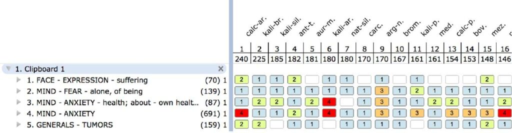
Plan: Calcarea arsenicosum LM1
Follow up one year later
It took her two months before she started the remedy because of a strong fear of an aggravation. Currently she was feeling good with lots of energy, especially in her business. She was able to relax easily. She was getting a lot of compliments on her skin since taking the medication. She no longer feared being robbed. Her fears at night had greatly improved. She was happier and more relaxed. Her vision had improved two lines in the right eye on the chart.
Physical Exam: Vision: 5/60 (2 lines better) in the right eye, 5/10 (no change) in the left eye. There were no demonstrable changes in the pigmentary findings in the right and left eye. Her blood pressure was more stable although there was no change in her medication. There was little change in retinal pigmentary findings, but since visual acuity is more closely associated with function of the retinal photoreceptor cells, it can be assumed that the change was due to improvement of function.
Case 2
RR, 78-year-old female with chief complaint of ARMD, “dry type,” cataracts and a “balance problem.”
She noticed her vision decreasing since her last eye exam when she was told that she had macular degeneration and cataracts. She complained of dim vision and trouble reading. She had episodes of vertigo that came on suddenly. “When they hit I go down like a rock.” With the vertigo she noticed that the room spun from left to right and she felt weak. She said she felt like a “quivering old lady.” She was so weak that her arms and legs shook. The vertigo occurred twice at 7:00 a.m. on waking. She pulled herself up with great effort. She lived in constant fear that an attack would occur when she was under stress. She was a real estate broker and, when she was in a stressful conversation, she could feel her balance leave her. She felt as if “the bottom is dropping out of my stomach and I feel light-headed.” She also had ringing in her ears along with the vertigo.
An important aspect of her life had always been ballroom dancing and she felt depressed now that she could no longer compete because of her condition. Her sexual drive had never been very high. Though not currently in a relationship, she felt a lot of sexuality when she danced. She especially liked the provocative aspects of Latin dance.
Physical exam: Vision: 20/300 right eye, 3/100 left eye. Advanced cataracts in both eyes. Myopic degeneration with atrophic changes in her retina. (Myopic degeneration is a type of macular degeneration associated with myopia. In this condition the retina is stretched and leads to an increase incidence of macular degeneration.)
Diagnosis: ARMD, Vertigo, Cataracts.
Rubrics
EYE: CATARACT
VERTIGO: ACCOMPANIED by, staggering
VERTIGO: SUDDEN
EAR: NOISES in, ringing
MIND: DANCING
FEMALE GENITALIA/SEX: SEXUAL DESIRE, diminished
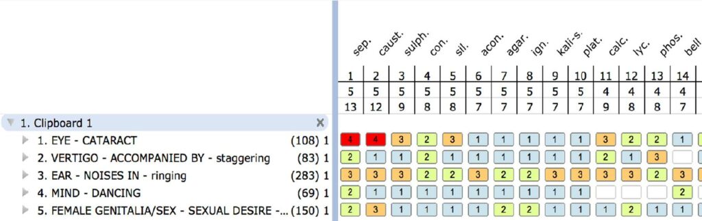
According to A.B. Norton in his article The Homeopathic Treatment of Incipient Senile Cataract, with Tabulated Results of One Hundred Cases, the number one homeopathic medicine to think of in females with advanced cataracts is Sepia.
Plan: Sepia LM1
Eight month follow up
She could read a menu. Her vision was 70 percent improved. Street signs were clearer and colors more vivid. There had been no more dizziness or episodes of vertigo. She had more strength and was more confident when walking. The ringing in her ears had gone. On PE, her vision was 20/200 in the right eye, a one line improvement. Her left eye showed a marked improvement from 20/200 to 3/100.
Discussion
At the eight-month follow-up there was a marked improvement of acuity although there was very little change in the density of the cataracts and the pigmentary changes. This would indicate that the remedy had had a positive effect on the function of the visual system.
Case 3
EA, 78-year-old female.
Two years ago, she developed cloudy vision in the left eye and letters began to run together while she was reading. One of the biggest difficulties in her life was her relationship with her daughter. “She has cut me off from the family. She will not return my letters and she will hang up the phone when I call. It is such a big disappointment in my life. She has told lies about my husband and me. She said, ‘You know mother, you are a slob.’ I wanted to cry, but I could not. How could she be so hurtful? We could go for hours not talking to each other.
“My daughter does not appreciate anything that I did. I wanted to tell them to ‘go to hell,’ but I did not because I did not want to exacerbate it. I blow up and get angry very quickly. Sometimes a silly little thing will irritate me. I was eating my salad and they brought out the main course. It was cold and I became extremely upset.”
Physical exam: Retinal pigment atrophy in the left eye. Vision 10/10 right (normal), 1/70 left eye
The rest of her physical exam was unremarkable and her blood pressure was normal.
Diagnosis: ARMD
Rubrics
MIND: CONTRADICTION; intolerant of contradiction
MIND: ANGER; trifles; at
MIND: ANGER; violent
CHEST: PALPITATION of heart, anger
VISION: RUN together, letters
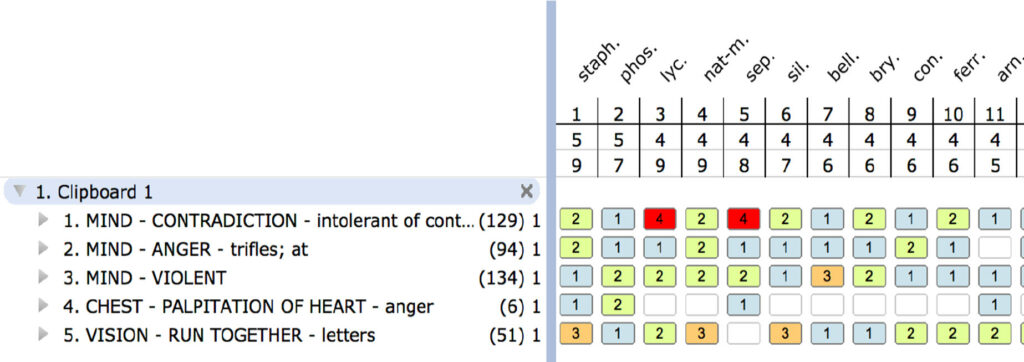
Plan: Staphysagria LM1
Seven month follow up
Peripheral vision was better in the left eye. She noticed an overall improvement in the right eye. Colors were much brighter; she owned a dress that she thought was gray and now it looked lavender! She was not getting angry as easily or as often. She called her daughter and asked her if she could meet with her and her daughter agreed. It was a good meeting and at the end she said that they both agreed they had wasted three years.
On PE, vision improved three lines in the left eye to 1/40 from 1/70 and 10/10 (unchanged) in the right eye.
Discussion
Age-related macular degeneration (ARMD) is the most common cause of irreversible blindness in people over 65 years of age. Risk factors include smoking, which increases the incidence of macular degeneration six fold, cardiovascular disease, hypertension, obesity, diabetes and nutritional deficiencies, especially of the antioxidants, zinc and vitamin D.(2)
Patients usually complain of blurred vision and difficulty with close work. They can also develop wavy lines and distortion of linear targets, a loss of color sense and the development of scotomas—small areas of blindness.
ARMD has become more prevalent in the world due to the above risk factors which may be associated with a general decline in nutrition and an increase in exposure to pesticides and preservatives, which increase the toxic load on the body. Heavy metals, in particular lead, cadmium and mercury, may all increase the risk of macular degeneration.(3)
From a homeopathic perspective, suppression of disease symptoms with allopathic treatment might also be a contributing factor. The overuse of antibiotics that weaken the immune system via the destruction of the gut microbiome have contributed to the alarming increase of inflammation and chronic diseases. Data from the18 year Speedwell eye study in the United Kingdom (4) showed that higher blood levels of C-reactive protein (an inflammatory marker) were associated with a higher risk of developing AMD. The homeopathic principle of suppression may also explain why cataract surgery leads to an increased incidence of macular degeneration. For example, the 5-year results of the Beaver Dam Eye Study (5) and the Blue Mountains Eye Study (6) identified persons with and without a history of cataract extraction at a baseline examination and reexamined them for the incidence of AMD at five and ten years. These studies found an association between cataract surgery and an increased 5-year incidence of late AMD. For the Beaver Dam Eye Study, cataract surgery before baseline was associated with an increased risk of advanced AMD and the Blue Mountains Eye Study showed a three-fold increased risk of advanced AMD.
Conventional, allopathic western ophthalmology explains this increase as due to the trauma of surgery or simply the natural progression of the aging process. However, homeopathic medicinal principles of suppression might also explain this association between cataract surgery and an increase risk of ARMD: when the underlying cause of disease (cataracts) is not treated, simply removing them surgically merely leads to the expression of the disease process in another organ or tissue.
There are currently no effective conventional treatments for the dry form of macular degeneration and pharmaceutical treatments for the wet form are limited by their toxicity and side effects. Steroid eye drops, for example, are known to increase the risk of cataracts and glaucoma. The treatment of wet macular degeneration with anti-VEGF (vascular endothelial growth factor) injections results in severe pathological changes that limit its use. Endophthalmitis, which is a severe infection that usually leads to blindness, can occur, as can retinal detachment. Glaucoma, as well as cataracts, can develop. A recent national study designed to compare the relative effectiveness of two frequently prescribed anti-VEGF medications to treat wet macular degeneration had some alarming results when it determined that, for more than 18% of those receiving the anti-VEGF treatment, the treatment itself was found to produce retinal geographic atrophy (GA), which is a more severe form of macular degeneration involving retinal cell death. The team of researchers led by Juan E. Grunwald, MD, of the University of Pennsylvania, has published a study of 1024 patients whose color fundus photos or fluorescein angiograms showed no visible signs of GA at enrollment. After two years, the researchers found that GA had developed in 187 patients, or 18.3% of the cohort being studied. They concluded that “anti-VEGF therapy may have a role in the development of GA.”
The homeopathic treatment of eye disease and particular macular degeneration is not new. New York Ophthalmic Hospital, which was founded in 1852, was under homeopathic management in 1867. In 1931 the hospital treated over 31,000 patients using a combination of a homeopathy and surgery. From 1879 to 1939 they offered Postgraduate courses in Homeopathic Ophthalmology. During this period, ophthalmology embraced homeopathic treatment. There were over ten textbooks on the subject (see References), a journal called Homeopathic Eye, Ear and Throat Journal (1895-1905), and a society called the American Homeopathic Ophthalmology and Otology Society (1877).
What makes homeopathy unique is the holistic treatment of macular degeneration that involves treating the whole person together with the pathology. When looking at a patient with AMD, the mental and emotional symptoms are just as important as the particular physical characteristics in the selection of the correct homeopathic medicine. All other contributing factors (patient’s medical history, environmental exposures, etc.) and the timeline of disease are documented. The homeopathic medicine is selected based on characteristic eye symptoms as well as the uniquely characteristic mental, emotional and physical symptoms of the patient. It is not the generic or pathognomonic symptoms of AMD that are used, but the peculiar, individualizing symptoms.
Other homeopathic remedies to consider for macular degeneration
The following tissue salts have been helpful in the treatment of ARMD:
- Calcarea flourica 8X – The tissue strengthener.
- Calcarea phosphorica 6X – The cell builder.
- Kali phosphorica 6X – Nerve nutrient.
- Natrum muriaticum 6X – The fluid distributor for dryness or excessive moisture in any part of the body. Can be especially helpful in cases of wet macular degeneration.
- Carboneum sulphuratum – Andrew Lange, ND, has reported success in early macular degeneration using Carboneum sulphuratum. He uses 30C daily and has seen resolution of drusen and pigmentary changes.
- Secale – Dr. Johann A. Miller has had good success with Secale in the treatment of macular degeneration. Secale is used in low potency when the patients have some general signs compatible with Secale, such as being worse from heat and better from cold.
- Sanicula and Vanadium – Dr. A.U. Ramakrishnan has used Sanicula in cases of macular degeneration with the symptom of wavy vision. He has also used Vanadium 200C monthly for the treatment of macular degeneration.
Sarcodes are homeopathic medicines prepared from actual tissue. Human retina has been used by Max Tetau in the 5C, 7C and 9C potencies. 5C appears to stimulate activity of the tissue from which it was derived. 7C appears to normalize tissue activity and 9C suppresses tissue activity.
Human retina 5C has been used with some success in stimulating the activity of the failing macula.
Homoeopathic Therapeutics in Ophthalmology by John L. Moffat, Ithaca, N. Y., 1916 lists the following remedies for macular degeneration:
For Neuroretinitis and Hyperemia: Bell., Dub., Gels.,Phos., Acon., Ars., Aur., Bry., Cact., Chin-s., Con., Fe-ph., Kail-i., Kali-m., Lach., Merc., Naph., Nux., Puls., Sec., Spig., Sulph., Ver-v.
Hemorrhagic: Arn., Bell., Crotalus horr.., Dub., Lach., Merc-cor., Phos
Pigmentosa: Lyc., Nux., Phos.
Punctata albescens: Naph.
Syphilitic: Asa., Aur., Kali-i., Merc-cor., Sol., Nit-ac.
The above homeopathic medicines and their indications were found during provings (homeopathy’s drug trials) or from the observation of accidental poisonings. One of the most important laws of homeopathy is the Law of Similars, which states that a substance that produces symptoms in a healthy person can treat those same symptoms when they occur in a sick person. In macular degeneration, we look for homeopathic medicines that will produce in healthy subjects the characteristic findings of the disease we are treating. For example, a toxic reaction or side effect of Carboneum sulphuratum and Plaquenil is a characteristic degeneration of the macula. Therefore, based on the Law of Similars, these homeopathic medicines, along with others that are listed in the vast homeopathic literature, are medicines to consider in the treatment of macular degeneration. In addition to the physical presentations of each remedy, there are also the characteristic mental and emotional symptoms. Looking at the totality of physical, mental and emotional symptoms leads to the proper homeopathic medicine prescription.
The three cases described in this report illustrate the benefits of homeopathic treatment in chronic macular degeneration, a condition that is therapeutically limited with conventional allopathic medicine. I remain hopeful that eye doctors will some day take a more open view of the homeopathic approach, especially in those cases that do not respond to allopathic treatment or where no treatment is available.
References
- www.ncbi.nlm.nih.gov/pmc/articles/PMC5178091/
- Ophthalmology 2001 Apr;108 (4):697-704. Risk factors for age-related macular degeneration: Pooled findings from three continents. Smith W1, Assink J, Klein R, et al.
- Ophthalmology. 2015 Jan;122 (1):129-37. Five heavy metallic elements and age-related macular degeneration: Korean National Health and Nutrition Examination Survey, 2008-2011.
- www.ncbi.nlm.nih.gov/pmc/articles/PMC3178119/
- Klein BE, Howard KP, Lee KE, et al. The relationship of cataract and cataract extraction to age-related macular degeneration: the Beaver Dam Eye Study. Ophthalmology. 2012;119(8):1628-1633.
- www.ncbi.nlm.nih.gov/pubmed/16935334
Other Resources
Kondrot EC. Healing the Eye the Natural Way: Alternative Medicine and Macular Degeneration. Carson City, NV: Nutritional Research Press; 2001.
Goldberg J, Flowerdew G, Smith E, Brody JA, Tso MO. Factors associated with age-related macular degeneration: an analysis of data from the first National Health and Nutrition Examination Survey. Am J Epidemiol. 1988;128(4):700-710.
Michael LD, Allen MJ. Nutritional supplementation, electrical stimulation and age related macular degeneration. J Orthomol Med. 1993;8(3):168-171.
Kondrot EC. Ten Essentials to Save Your Sight. Charleston, SC: Advantage; 2012.
Grunwald JE, Pistilli M, Ying GS, Maguire MG, et al. Comparison of Agerelated Macular Degeneration Treatments Trials. Ophthalmology. 2015 Apr; 122(4):809-816.
Blair CJ, Ferguson J, Jr. Exacerbation of senile macular degeneration following cataract extraction. Am J Ophthalmol. 1979;87(1):77-83.
Angell, H.C. Treatise on the Diseases of the Eye. 1871
Becker, A. Diseases of the Eye, Treated Homeopathically. Boyle, C. Therapeutics of the Eye
Berridge, E.W. Complete Repertory to the Homeopathic Materia Medica Diseases of the Eye, 1873
Buffum, J. Diseases & Injuries of the Eye. 1883
Buffum, J Manual of the Essentials of the Diseases of the Eye & Ear. 1896
Moffat, J. Homeopathic Therapeutics in Ophthalmology. 1916 1916
McBride, N. Diseases of the Eye 1897
Norton, G. and Allen, T. F. Ophthalmic Therapeutics 1876
Norton, A. B. Ophthalmic Diseases & Therapeutics 1872
Norton, A.B. The Essentials of the Diseases of the Eye. 1904
Peters, J. A. A Treatise on the Diseases of the Eye. 1854
Levy T. Curing the Incurable: Vitamin C, Infectious Diseases, and Toxins. Philadelphia, PA: Xlibris Corp; 2002.
Schaumberg DA, Mendes F, Balaram M, Dana MR, Sparrow D, Hu H. Accumulated lead exposure and risk of age-related cataract in men. JAMA. 2004;292(22):2750-2754.
About the author: Ed Kondrot, MD, CCH, DHt, has practiced ophthalmology for over 20 years and classical homeopathy for over 15 years. He established a healing center, Healing the Eye, located in Zephyrhills, Florida, which treats patients who seek holistic non-invasive therapies for eye problems that include macular degeneration, glaucoma, cataracts, eyestrain and other eye disorders. He is board certified in ophthalmology, certified by the Council for Homeopathic Certification and is a diplomate of the American Board of Homeopathic Medicine. He has authored several books and hosts the weekly radio show Healthy Vision Talk Radio broadcasted on KFNX Talk Radiohosts.